Four single-cell analysis technologies go head-to-head
The rise of the spatial omics market is demonstrated by the variety of available platforms, which provide different kinds of information and use different samples, such as suspension, bulk, RNA or protein.

Spatial biology is an exciting frontier. Moving from bulk analysis to single-cell resolution means you can now see where signal is coming from and what cell types are contributing. Spatial information provides a new layer of details: where cells are located in the tissue, how they’re interacting and what they’re interacting with. You’re also able to ask new questions and better understand the biology at hand.
During Session I of our latest IMC™ Forum, Stepping Through the Pipeline: Recommendations and Considerations for In-Depth Analysis, Won Jin Ho, MD, Assistant Professor at the Sidney Kimmel Comprehensive Cancer Center at Johns Hopkins University, gives us perspective on how Imaging Mass Cytometry™ fits into the range of omics platforms.
The rise of the spatial omics market is demonstrated by the variety of available platforms. Researchers are able to compare by dimensionality, how many parameters you can look at in a given sample and how much the process costs. Different platforms provide different kinds of information and use different samples, such as suspension, bulk, RNA or protein.
Here are the pros and cons of four technologies:
Immunohistochemistry is a simple, low-plex and low-cost technology that can reliably look at 1–3 markers. You’re able to examine a large tissue area, and since it’s light microscopy-based, researchers are able to appreciate morphology. However, you can only see a few markers, so it takes significant resources to attain the insights higher-plex imaging would provide.
Bulk transcriptomics allows you to look at tens of thousands of parameters with a low cost, but you aren’t able to get any spatial information.
Spatial transcriptomics also allows you to look at tens of thousands of parameters, but it’s costly and has limited cellular resolution.
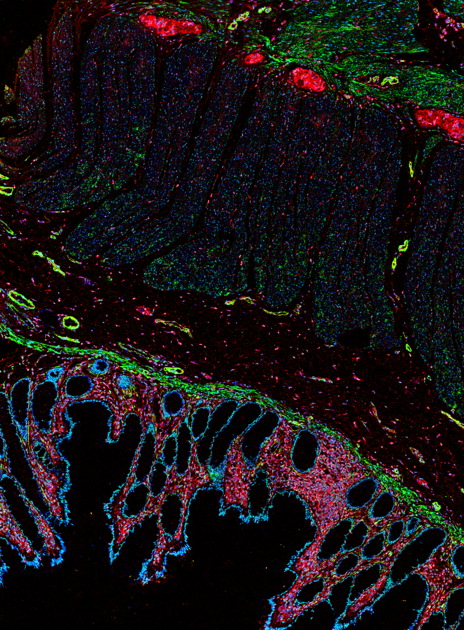
IMC is based on IHC, but because it uses metal reporters, you don’t have to worry about autofluorescence. This is an important factor in research work for particular cancers in which tissues are highly autofluorescent. IMC readouts are also more quantitative than fluorescent technologies.
Standardized solutions to get you started or accelerate your current IMC research
- The IMC Cell Segmentation Kit (PN TIS-00001) performs segmentation based on plasma membrane and can be added to nearly any panel without modifications.
- Our Human and Mouse Immuno-Oncology IMC Panel Kits are verified mix-and-match panel kits to standardize your workflow. They enable researchers to quickly develop a deep understanding of the tumor microenvironment and can be easily supplemented with catalog antibodies.
Check out other blog posts
World Diabetes Day

Where should managers put their focus to ensure their labs run smoothly? Learn about a new metric that can help improve lab efficiencies and cost savings for years to come.
Scaling up and keeping pace

Are you asking the right question? Considerations for when to automate, from common drivers to what you really should be thinking about.
ASHG 2022

Luke Stewart, Director, Field Applications, and two special guests will be leading a workshop on biobank workstream processes at ISBER's 2022 Annual Meeting.
Unless explicitly and expressly stated otherwise, all products are provided for Research Use Only, not for use in diagnostic procedures. Find more information here.